Hoof trimming is not a pedicure—it is biomechanical engineering. Every stroke of the knife or grinder alters the distribution of ground reaction forces acting on the bovine limb. While various schools of thought are emerging globally (e.g., the White Line Atlas method popular in the USA), in Europe and the majority of professional operations, the undisputed foundation remains the Dutch Method, developed by Dr. Egbert Toussaint Raven.
Understanding this method does not rely on the mechanical rote memorization of 5 steps, but rather on comprehending why each step is performed.
Objective: Restoring Balance
The dairy cow, particularly the Holstein-Friesian (HF) breed, exhibits a natural tendency toward overgrowth of the lateral claw in the hindlimbs (pelvic limbs) and the medial claw in the forelimbs (thoracic limbs). This leads to overloading of the respective claw, damage to the corium, and ulcer formation. The objective of trimming is to restore the natural conformation and ensure even weight distribution across both claws.
The 5-Step Method by Toussaint Raven
This method is divided into two stages: Functional Trimming (Steps 1–3), performed on every cow, and Therapeutic/Corrective Trimming (Steps 4–5), applied only when pathological lesions are identified.
Step 1: Establishing Length and Thickness (The Model Claw)
We begin with the stable claw. In the hindlimbs, this is the medial claw (it bears less weight and typically retains a more natural shape).
- Length: We measure the dorsal wall length from the coronary band to the toe tip. For an adult HF cow, the standard is 7.5 cm. Anything extending beyond this dimension is trimmed perpendicular to the sole.
- Note: In heifers or smaller breeds (Jerseys), this dimension may be 7 cm.
- Thickness: We trim the sole at the toe, leaving a minimal thickness of approx. 5–7 mm. In the posterior part of the sole (the heel), we aim to preserve as much horn as possible to maintain the correct claw angle.
- Principle: The sole should be flat and perpendicular to the long axis of the metatarsal bone.
Step 2: Matching the Overloaded Claw
We proceed to the lateral claw (hindlimb). This is typically the overgrown claw.
- We trim it to the same dorsal length as the medial claw (from Step 1).
- We reduce the sole level to match the level of the medial claw.
- Objective: When a flat surface (e.g., a knife) is applied across both claws, they should form a single, flat plane. This ensures equal weight bearing on both digits.
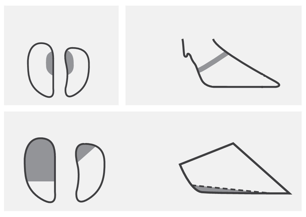
Step 3: Modeling (Dishing)
This is a critical moment for the prophylaxis of typical ulcers. A “dish” or “hollow” is excised along the axial (inner) margins of both claws (within the interdigital space).
- The dish should be gradual.
- Objective: To offload the typical site for Rusterholz ulcers (the insertion site of the deep digital flexor tendon) and to facilitate the self-cleaning of the interdigital space.
- Important: Do not compromise the weight-bearing wall (abaxial margin) or the toe tip!
If the claws are healthy, the procedure ends here. However, if lesions were identified during Steps 1–3, we proceed to the therapeutic stages.
Step 4: Offloading Diseased Tissues
If an ulcer, white line lesion, or hemorrhage is present on one claw (usually the lateral), we must transfer the weight to the healthy claw.
- We significantly lower the heel of the affected claw so that the healthy claw (medial) assumes the majority of the load.
- In cases of severe pain, we utilize orthopedic blocks (wood or rubber), adhered to the healthy claw. This is the most effective treatment for lameness, giving the affected claw a “vacation” from ground contact.
Step 5: Removing Loose Horn and Control
We remove all loose, undermined horn under which anaerobic bacteria may proliferate.
- Particular attention is paid to the heels (Digital Dermatitis) and the white line.
- Sharp edges of the excised lesions must be smoothed to prevent irritation of the corium.
Other Approaches and Modifications
Although the 5-Step Method is dominant, it is worth acknowledging other perspectives:
- White Line Atlas Method (USA): Focuses more on sole thickness determined by the visibility of the white line, rather than rigid adherence to the 7.5 cm dimension. This is a more advanced approach requiring significant experience in reading hoof horn, useful in herds with atypical horn wear (e.g., highly abrasive concrete).
- Pasture Trimming (New Zealand/Ireland): In systems where cows walk extensively on soft ground, trimming is less aggressive. It focuses mainly on shortening the toe, as heel wear is lower than in free-stall barns.
Summary
The Dutch Method is not merely a cutting technique—it is a herd health management strategy. Correctly performed functional trimming (Steps 1–3) is the best prevention. Conversely, precise offloading (Step 4) using blocks is the only path to healing sole ulcers.
Remember: “Horn grows slowly, but can disappear in a second.” Every movement of the knife must be deliberate.
Bibliography:
- Toussaint Raven, E. (1989). Cattle Footcare and Claw Trimming. Farming Press.
- Greenough, P. R., & Weaver, A. D. (1997). Lameness in Cattle. W.B. Saunders.
- Blowey, R. W. (2011). Cattle Lameness and Hoofcare: An Illustrated Guide. 5m Publishing.
- Shearer, J. K., & van Amstel, S. R. (2001). Functional and Corrective Claw Trimming. The Veterinary Clinics of North America. Food Animal Practice.
- Burgi, K. (Dairyland Hoof Care Institute). Functional Hoof Trimming & Hoof Health.
- Chesterton, R. N. (1989). Examination and trimming of the cow’s foot. Proceedings of the Society of Dairy Cattle Veterinarians of the NZVA.
Additional Training Materials:
- Zinpro Corp. Cattle Lameness: Identification, Prevention and Control of Claw Lesions. (Excellent visual atlas of lesions and anatomy).
- University of Wisconsin-Madison (School of Veterinary Medicine): The Dairyland Initiative – Hoof Health Supplement.